Home
Uncategories
Compact Bone Diagram - Long Bone Anatomy Diaphysis Shaft Of The Bone Made Of Compact Bone Ppt Video Online Download / Compact bone is formed from a number of osteons, which are circular units of bone material and blood vessels.
Compact Bone Diagram - Long Bone Anatomy Diaphysis Shaft Of The Bone Made Of Compact Bone Ppt Video Online Download / Compact bone is formed from a number of osteons, which are circular units of bone material and blood vessels.
Compact Bone Diagram - Long Bone Anatomy Diaphysis Shaft Of The Bone Made Of Compact Bone Ppt Video Online Download / Compact bone is formed from a number of osteons, which are circular units of bone material and blood vessels.. Haversian canals (sometimes canals of havers) are a series of microscopic tubes in the outermost region of bone called cortical bone. Thin layer of reticular ct lining internal marrow cavity. There are pores and spaces even in compact bone. It is also called osseous tissue or cortical bone and it provides structure and support for an organism as part of its skeleton, in addition to being a location for the storage of minerals like calcium.about 80% of the weight of the human skeleton comes from. Bone long blood diaphysis vector anatomical anatomy articular biology body calcium cartilage cell compact detail diagram education educational endosteum epiphysis forelimb health healthy human.
There are small canals that run through the bone, which allow blood vessels to penetrate it. Compact bone tissue diagram quizlet. You need to get 100% to score the 15 points available. There are pores and spaces even in compact bone. Compact bone is the denser, stronger of the two types of osseous tissue (figure 6.3.6).
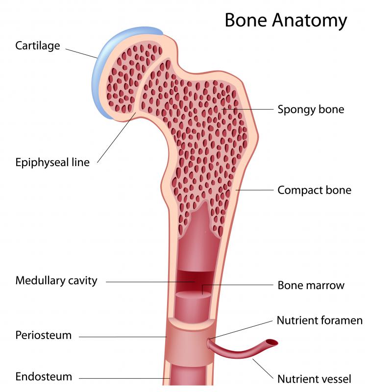
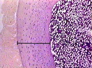
What Is Compact Bone With Pictures from images.infobloom.com Online quiz to learn compact bone diagram; A diagram of the anatomy of a bone, showing the compact bone. Compact bone is the strongest form of bone tissue containing few spaces. Bone marrow diagram, compact bone diagram quiz, compact bone slide labeled, diagram long bone, labeled compact bone model. Compact bone, also called cortical bone, dense bone in which the bony matrix is solidly filled with organic ground substance and inorganic salts, leaving only tiny spaces (lacunae) that contain the osteocytes, or bone cells.compact bone makes up 80 percent of the human skeleton; Please like this video share with all the learners comment your opinion and subscribe to support my channel this is a small step to teach what i know to. (b) in this micrograph of the osteon, you can clearly see the concentric lamellae and central canals. In long bones, as you move from the outer cortical compact bone to the inner medullary cavity, the bone transitions to spongy bone.
Online quiz to learn compact bone diagram;
Compact bone diagram osteon compact bone ap pinterest anatomy human anatomy and. Cancellous bones, compact bone, cortical bone, diaphyses, haversian canal. As seen in the image below, compact bone forms the cortex, or hard outer shell of most bones in the body. Provides protection and support while resisting stress from weight and movement. (b) in this micrograph of the osteon, you can clearly see the concentric lamellae and central canals. Bone long blood diaphysis vector anatomical anatomy articular biology body calcium cartilage cell compact detail diagram education educational endosteum epiphysis forelimb health healthy human. Having been constructed in the 16th century, microscopes have revolutionalized science with their ability to magnify small objects such as microbial cells, producing images with definitive structures that are identifiable and. The remainder of the bone is formed by cancellous or spongy bone. It makes up the outer cortex of all bones and is in immediate contact with the periosteum. Compact bone tissue diagram quizlet. Pig bone diagram wiring diagram, femur bone diagram full human skeleton diagram femur simple anatomy, colored ear diagram for kids bone labeled of the eye to label compact bone diagram simple diagram system. Because of its strength, the compact bone makes it possible for the bone to support weight. (b) in this micrograph of the osteon, you can clearly see the concentric lamellae and central canals.
Compact bone is formed from a number of osteons, which are circular units of bone material and blood vessels. Provides protection and support while resisting stress from weight and movement. A diagram of the anatomy of a bone, showing the compact bone. There are two types of bone tissue: Compact and spongy.the names imply that the two types differ in density, or how tightly the tissue is packed together.
Bone Compact Decalcified C S from www.austincc.edu In long bones, as you move from the outer cortical compact bone to the inner medullary cavity, the bone transitions to spongy bone. The remainder is cancellous bone, which has a spongelike appearance with numerous large spaces and is found in the. Online quiz to learn compact bone diagram; Skincare korea untuk remaja smp. Thin layer of reticular ct lining internal marrow cavity. Anatomy of rib cage 12 photos of the anatomy of rib cage anatomical rib cage necklace, anatomy and physiology of rib cage, anatomy of human rib cage, anatomy of rib cage area, human anatomy rib cage muscles, human anatomy, anatomical rib cage necklace, anatomy and physiology of rib cage, anatomy of human rib cage, anatomy … Haversian canals (sometimes canals of havers) are a series of microscopic tubes in the outermost region of bone called cortical bone. Compact bone is the strongest form of bone tissue containing few spaces.
There are pores and spaces even in compact bone.
Anatomy of rib cage 12 photos of the anatomy of rib cage anatomical rib cage necklace, anatomy and physiology of rib cage, anatomy of human rib cage, anatomy of rib cage area, human anatomy rib cage muscles, human anatomy, anatomical rib cage necklace, anatomy and physiology of rib cage, anatomy of human rib cage, anatomy … There are pores and spaces even in compact bone. Please like this video share with all the learners comment your opinion and subscribe to support my channel this is a small step to teach what i know to. Compact and spongy.the names imply that the two types differ in density, or how tightly the tissue is packed together. Thin layer of reticular ct lining internal marrow cavity. 850 x 560 png 177 кб. Cancellous bones, compact bone, cortical bone, diaphyses, haversian canal. Nov diagram for.net is a fully managed, extensible and powerful diagramming framework, which can help you create feature rich diagramming solutions in winforms, wpf, silverlight, xamarin.mac, monomac and asp. (b) in this micrograph of the osteon, you can clearly see the concentric lamellae and central canals. Pig bone diagram wiring diagram, femur bone diagram full human skeleton diagram femur simple anatomy, colored ear diagram for kids bone labeled of the eye to label compact bone diagram simple diagram system. It is dense (because of calcified matrix) with tiny spaces known as lucanas. Related posts of compact bone diagram labeled anatomy of rib cage. Online quiz to learn compact bone diagram;
Learn vocabulary, terms, and more with flashcards, games, and other study tools. It makes up the outer cortex of all bones and is in immediate contact with the periosteum. Anatomy of rib cage 12 photos of the anatomy of rib cage anatomical rib cage necklace, anatomy and physiology of rib cage, anatomy of human rib cage, anatomy of rib cage area, human anatomy rib cage muscles, human anatomy, anatomical rib cage necklace, anatomy and physiology of rib cage, anatomy of human rib cage, anatomy … The outer part of a long bone is made of compact bone. A diagram of the anatomy of a bone, showing the compact bone.
1 from Compact bone is the strongest form of bone tissue containing few spaces. About press copyright contact us creators advertise developers terms privacy policy & safety how youtube works test new features press copyright contact us creators. The remainder is cancellous bone, which has a spongelike appearance with numerous large spaces and is found in the. Thin layer of reticular ct lining internal marrow cavity. Human bone generally comprises osseous tissue, an outer coating called a periosteum, and bone marrow.the two main structural components typically include spongy bone on the interior, with an outer layer of compact bone. Compact bone diagram osteon compact bone ap pinterest anatomy human anatomy and. All of these tissue types are shown in figure below. The remainder of the bone is formed by cancellous or spongy bone.
850 x 560 png 177 кб.
Cancellous bones, compact bone, cortical bone, diaphyses, haversian canal. Compact bone, also called cortical bone, dense bone in which the bony matrix is solidly filled with organic ground substance and inorganic salts, leaving only tiny spaces (lacunae) that contain the osteocytes, or bone cells.compact bone makes up 80 percent of the human skeleton; Compact bone diagram osteon compact bone ap pinterest anatomy human anatomy and. Usually found in long bones of the body, it consists of units called. Usually bones that are thin and curved. Although the calls are close together, this type of bone is not completely solid. Compact and spongy.the names imply that the two types differ in density, or how tightly the tissue is packed together. Related posts of compact bone diagram labeled abdominal organs kidney. It is dense (because of calcified matrix) with tiny spaces known as lucanas. A diagram of the anatomy of a bone, showing the compact bone. Human bone generally comprises osseous tissue, an outer coating called a periosteum, and bone marrow. Online quiz to learn compact bone diagram; Learn vocabulary, terms, and more with flashcards, games, and other study tools.
0 Comments:
Posting Komentar